Abstract
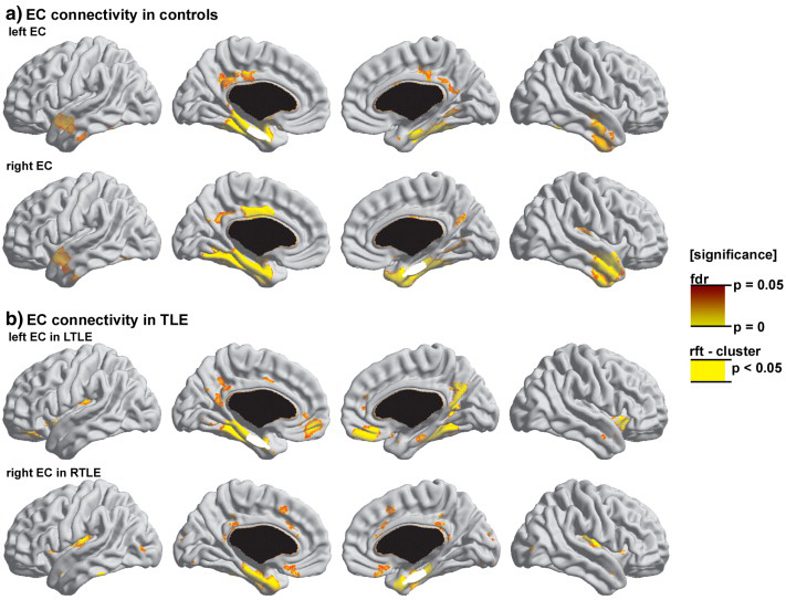
Temporal lobe epilepsy (TLE) is considered primarily a limbic disorder. Our purpose was to map limbic network organization in TLE and to statistically relate it to neocortical atrophy. We performed MRI-based cortical thickness analysis in 110 TLE patients (including 68 patients with hippocampal atrophy and 42 patients with normal hippocampal volume) and 46 healthy controls. Limbic connectivity was statistically inferred by correlating mean thickness of the entorhinal cortex (EC) with thickness at each vertex across the entire neocortex. The EC was chosen as seed region since it is the link between the neocortex and the hippocampal formation. Patients showed cortical thinning mainly in temporal and fronto-central neocortices, with a prevalence of atrophy in up to 35%. In controls, EC networks corresponded closely to known anatomical connections. In TLE the pattern of correlations was similar to controls, suggesting that pathological processes in the EC affect the same networks that co-vary with the EC in the healthy brain. Nevertheless, we found decreases in correlations mainly in the temporal lobe and increases mainly in orbitofrontal cortices. Although our analysis indicated alterations in the temporo-limbic network in TLE, there was no association between mesiotemporal connectivity and atrophy across the entire cortical surface. This divergence underlines the complexity of the pathophysiological mechanisms leading to neocortical atrophy in TLE.