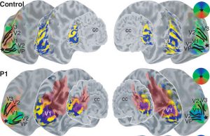
Abstract
Polymicrogyrias (PMG) are cortical malformations resulting from developmental abnormalities. In animal models PMG has been associated with abnormal anatomy, function, and organization. The purpose of this study was to describe the function and organization of human polymicrogyric cortex using functional magnetic resonance imaging. Three patients with epilepsy and bilateral parasagittal occipital polymicrogyri were studied. They all had normal vision as tested by Humphrey visual field perimetry. The functional organization of the visual cortex was reconstructed using phase-encoded retinotopic mapping analysis. This method sequentially stimulates each point in the visual field along the axes of a polar-coordinate system, thereby reconstructing the representation of the visual field on the cortex. We found normal cortical responses and organization of early visual areas (V1, V2, and V3/VP). The locations of these visual areas overlapped substantially with the PMG. In five out of six hemispheres the reconstructed primary visual cortex completely fell within polymicrogyric areas. Our results suggest that human polymicrogyric cortex is not only organized in a normal fashion, but is also actively involved in processing of visual information and contributes to normal visual perception.
