Abstract
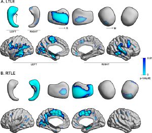
In drug-resistant temporal lobe epilepsy (TLE), MRI studies have shown consistent mesiotemporal and neocortical structural alterations when comparing patients to healthy controls. It remains, however, relatively unclear whether the side of seizure focus differentially impacts the degree of structural damage. This work performed a comprehensive surface-based analysis of mesiotemporal and neocortical morphology on preoperative 1.5 T MRI in 25/35 LTLE/RTLE patients that achieved seizure freedom after surgery (i.e., Engel-I outcome; 7 ± 2 years follow-up), an imaging-independent confirmation of focus lateralization. Compared to 46 age- and sex-matched controls, both TLE groups displayed marked ipsilateral atrophy in mesiotemporal regions, while cortical thinning was bilateral. Direct contrasts between LTLE and RTLE did not reveal significant differences. Bootstrap simulations indicated low reproducibility of observing a between-cohort difference; power analysis revealed that more than 110 patients would be necessary to detect subtle differences. No difference between LTLE and RTLE was confirmed when using voxel-based morphometry, an independent proxy of gray matter volume. Similar results were obtained analyzing a separate 3 T dataset (15/15 LTLE/RTLE patients; Engel-I after 4 ± 2 years follow-up; 42 controls). Our results strongly support equivalent gray matter compromise in left and right TLE. The morphological profile of seizure-free patients, presenting with ipsilateral mesiotemporal and bilateral cortical atrophy, motivates the development of neuromarkers of outcome that consider both mesiotemporal and neocortical structures.
