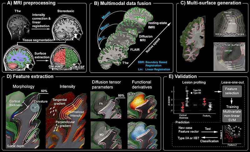
Abstract
Focal cortical dysplasia (FCD), a malformation of cortical development, is a frequent cause of drug-resistant epilepsy. This lesion is histologically classified into Type-IIA (dyslamination, dysmorphic neurons) and Type-IIB (dyslamination, dysmorphic neurons, and balloon cells). Reliable in-vivo identification of lesional subtypes is important for preoperative decision-making and surgical prognostics. We propose a novel multi-modal MRI lesion profiling based on multiple surfaces that systematically sample intra- and subcortical tissue. We applied this framework to histologically-verified FCD. We aggregated features describing morphology, intensity, microstructure, and function from T1-weighted, FLAIR, diffusion, and resting-state functional MRI. We observed alterations across multiple features in FCD Type-IIB, while anomalies in IIA were subtle and mainly restricted to FLAIR intensity and regional functional homogeneity. Anomalies in Type-IIB were seen across all intra- and sub-cortical levels, whereas those in Type-IIA clustered at the cortico-subcortical interface. A supervised classifier predicted the FCD subtype with 91% accuracy, validating our profiling framework at the individual level.